The skeletal systemis a complex network of bones and... Show more
Sign up to see the contentIt's free!
Access to all documents
Improve your grades
Join milions of students
Knowunity AI
Subjects
Triangle Congruence and Similarity Theorems
Triangle Properties and Classification
Linear Equations and Graphs
Geometric Angle Relationships
Trigonometric Functions and Identities
Equation Solving Techniques
Circle Geometry Fundamentals
Division Operations and Methods
Basic Differentiation Rules
Exponent and Logarithm Properties
Show all topics
Human Organ Systems
Reproductive Cell Cycles
Biological Sciences Subdisciplines
Cellular Energy Metabolism
Autotrophic Energy Processes
Inheritance Patterns and Principles
Biomolecular Structure and Organization
Cell Cycle and Division Mechanics
Cellular Organization and Development
Biological Structural Organization
Show all topics
Chemical Sciences and Applications
Atomic Structure and Composition
Molecular Electron Structure Representation
Atomic Electron Behavior
Matter Properties and Water
Mole Concept and Calculations
Gas Laws and Behavior
Periodic Table Organization
Chemical Thermodynamics Fundamentals
Chemical Bond Types and Properties
Show all topics
European Renaissance and Enlightenment
European Cultural Movements 800-1920
American Revolution Era 1763-1797
American Civil War 1861-1865
Global Imperial Systems
Mongol and Chinese Dynasties
U.S. Presidents and World Leaders
Historical Sources and Documentation
World Wars Era and Impact
World Religious Systems
Show all topics
Classic and Contemporary Novels
Literary Character Analysis
Rhetorical Theory and Practice
Classic Literary Narratives
Reading Analysis and Interpretation
Narrative Structure and Techniques
English Language Components
Influential English-Language Authors
Basic Sentence Structure
Narrative Voice and Perspective
Show all topics
841
•
Updated Mar 20, 2026
•
Reese Kirk
@reesekirk_qsqr
The skeletal systemis a complex network of bones and... Show more


Bone shape and function structure anatomy is intricately designed to support various bodily functions. The structure of bones is closely related to their specific roles within the skeletal system.
Long bones, such as those found in the forearms, have distinct parts:
Definition: Periosteum - A tough, fibrous membrane that covers the outer surface of bones, playing a crucial role in bone growth and repair.
Bone tissue function varies depending on its location and structure. Two main types of bone tissue are found in the skeletal system:
Compact bone (cortical bone):
Spongy bone (cancellous bone):
Highlight: The structure of bones is optimized for their specific functions, with compact bone providing strength and spongy bone offering flexibility and space for bone marrow.
The medullary cavity, a hollow chamber formed by compact bone in the diaphysis of long bones, is lined by a layer of cells called the endosteum and filled with marrow. This cavity plays a crucial role in bone metabolism and blood cell production.

The microscopic structure of bone is complex and highly organized, particularly in compact bone. The basic unit of compact bone is called an osteon, which consists of several key components:
Vocabulary: Osteon - The cylindrical unit of compact bone, also known as a Haversian system.
Perforating canals, also called Volkmann's canals, are transverse channels that connect central canals, facilitating communication and nutrient distribution throughout the bone tissue.
Spongy bone structure differs from compact bone in that it lacks the organized Haversian system. Instead, spongy bone cells lie within trabeculae and receive nutrients through diffusion into canaliculi.
Highlight: The microscopic structure of bone allows for efficient nutrient distribution and communication between bone cells, contributing to the overall health and function of the skeletal system.
Understanding the microscopic structure of bone is crucial for comprehending bone physiology, growth, and repair processes. This knowledge forms the basis for studying various bone-related conditions and developing treatments for skeletal disorders.

Bone development and growth, known as ossification or osteogenesis, is a complex process that begins during the first few weeks of prenatal development and continues into early adulthood. This process involves the formation and remodeling of bone tissue through the actions of specialized cells.
There are two main ways in which bones form by replacing existing connective tissue:
Definition: Ossification - The process of bone formation and development, also known as osteogenesis.
Key cells involved in bone development and growth include:
Endochondral bone development involves several stages:
Highlight: The epiphyseal plate, also known as the growth plate, is crucial for bone elongation during childhood and adolescence.
Ossification is not complete until early adulthood when the epiphyseal plates close, and bone growth ceases. Understanding the process of bone development and growth is essential for comprehending various skeletal disorders and developing treatments for bone-related conditions.

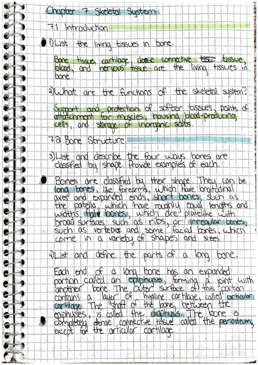
The skeletal system is a crucial component of human anatomy, comprising various living tissues in bone and serving multiple essential functions. This chapter explores the structure, development, and growth of bones, providing a comprehensive overview of the skeletal system.
Vocabulary: Ossification - The process of bone formation, also known as osteogenesis.
The skeletal system's functions include:
Highlight: The skeletal system is not just a static framework but a dynamic system that plays vital roles in various bodily functions.
Bone shape and function structure anatomy is diverse, with bones classified into four main types based on their shape:
Example: The forearm contains long bones, which have longitudinal axes and expanded ends, making them ideal for providing leverage and support for arm movements.
Understanding the structure and classification of bones is essential for comprehending their functions and roles within the body. This knowledge forms the foundation for further exploration of the skeletal system's complexities.


Our AI companion is specifically built for the needs of students. Based on the millions of content pieces we have on the platform we can provide truly meaningful and relevant answers to students. But its not only about answers, the companion is even more about guiding students through their daily learning challenges, with personalised study plans, quizzes or content pieces in the chat and 100% personalisation based on the students skills and developments.
You can download the app in the Google Play Store and in the Apple App Store.
That's right! Enjoy free access to study content, connect with fellow students, and get instant help – all at your fingertips.
App Store
Google Play
The app is very easy to use and well designed. I have found everything I was looking for so far and have been able to learn a lot from the presentations! I will definitely use the app for a class assignment! And of course it also helps a lot as an inspiration.
Stefan S
iOS user
This app is really great. There are so many study notes and help [...]. My problem subject is French, for example, and the app has so many options for help. Thanks to this app, I have improved my French. I would recommend it to anyone.
Samantha Klich
Android user
Wow, I am really amazed. I just tried the app because I've seen it advertised many times and was absolutely stunned. This app is THE HELP you want for school and above all, it offers so many things, such as workouts and fact sheets, which have been VERY helpful to me personally.
Anna
iOS user
I think it’s very much worth it and you’ll end up using it a lot once you get the hang of it and even after looking at others notes you can still ask your Artificial intelligence buddy the question and ask to simplify it if you still don’t get it!!! In the end I think it’s worth it 😊👍 ⚠️Also DID I MENTION ITS FREEE YOU DON’T HAVE TO PAY FOR ANYTHING AND STILL GET YOUR GRADES IN PERFECTLY❗️❗️⚠️
Thomas R
iOS user
Knowunity is the BEST app I’ve used in a minute. This is not an ai review or anything this is genuinely coming from a 7th grade student (I know 2011 im young) but dude this app is a 10/10 i have maintained a 3.8 gpa and have plenty of time for gaming. I love it and my mom is just happy I got good grades
Brad T
Android user
Not only did it help me find the answer but it also showed me alternative ways to solve it. I was horrible in math and science but now I have an a in both subjects. Thanks for the help🤍🤍
David K
iOS user
The app's just great! All I have to do is enter the topic in the search bar and I get the response real fast. I don't have to watch 10 YouTube videos to understand something, so I'm saving my time. Highly recommended!
Sudenaz Ocak
Android user
In school I was really bad at maths but thanks to the app, I am doing better now. I am so grateful that you made the app.
Greenlight Bonnie
Android user
I found this app a couple years ago and it has only gotten better since then. I really love it because it can help with written questions and photo questions. Also, it can find study guides that other people have made as well as flashcard sets and practice tests. The free version is also amazing for students who might not be able to afford it. Would 100% recommend
Aubrey
iOS user
Best app if you're in Highschool or Junior high. I have been using this app for 2 school years and it's the best, it's good if you don't have anyone to help you with school work.😋🩷🎀
Marco B
iOS user
THE QUIZES AND FLASHCARDS ARE SO USEFUL AND I LOVE Knowunity AI. IT ALSO IS LITREALLY LIKE CHATGPT BUT SMARTER!! HELPED ME WITH MY MASCARA PROBLEMS TOO!! AS WELL AS MY REAL SUBJECTS ! DUHHH 😍😁😲🤑💗✨🎀😮
Elisha
iOS user
This app is phenomenal down to the correct info and the various topics you can study! I greatly recommend it for people who struggle with procrastination and those who need homework help. It has been perfectly accurate for world 1 history as far as I’ve seen! Geometry too!
Paul T
iOS user
Reese Kirk
@reesekirk_qsqr
The skeletal system is a complex network of bones and tissues that provides support, protection, and various functions for the human body. This summary explores the structure, development, and growth of bones, highlighting key concepts in bone anatomy and physiology.... Show more
Access to all documents
Improve your grades
Join milions of students
Bone shape and function structure anatomy is intricately designed to support various bodily functions. The structure of bones is closely related to their specific roles within the skeletal system.
Long bones, such as those found in the forearms, have distinct parts:
Definition: Periosteum - A tough, fibrous membrane that covers the outer surface of bones, playing a crucial role in bone growth and repair.
Bone tissue function varies depending on its location and structure. Two main types of bone tissue are found in the skeletal system:
Compact bone (cortical bone):
Spongy bone (cancellous bone):
Highlight: The structure of bones is optimized for their specific functions, with compact bone providing strength and spongy bone offering flexibility and space for bone marrow.
The medullary cavity, a hollow chamber formed by compact bone in the diaphysis of long bones, is lined by a layer of cells called the endosteum and filled with marrow. This cavity plays a crucial role in bone metabolism and blood cell production.
Access to all documents
Improve your grades
Join milions of students
The microscopic structure of bone is complex and highly organized, particularly in compact bone. The basic unit of compact bone is called an osteon, which consists of several key components:
Vocabulary: Osteon - The cylindrical unit of compact bone, also known as a Haversian system.
Perforating canals, also called Volkmann's canals, are transverse channels that connect central canals, facilitating communication and nutrient distribution throughout the bone tissue.
Spongy bone structure differs from compact bone in that it lacks the organized Haversian system. Instead, spongy bone cells lie within trabeculae and receive nutrients through diffusion into canaliculi.
Highlight: The microscopic structure of bone allows for efficient nutrient distribution and communication between bone cells, contributing to the overall health and function of the skeletal system.
Understanding the microscopic structure of bone is crucial for comprehending bone physiology, growth, and repair processes. This knowledge forms the basis for studying various bone-related conditions and developing treatments for skeletal disorders.
Access to all documents
Improve your grades
Join milions of students
Bone development and growth, known as ossification or osteogenesis, is a complex process that begins during the first few weeks of prenatal development and continues into early adulthood. This process involves the formation and remodeling of bone tissue through the actions of specialized cells.
There are two main ways in which bones form by replacing existing connective tissue:
Definition: Ossification - The process of bone formation and development, also known as osteogenesis.
Key cells involved in bone development and growth include:
Endochondral bone development involves several stages:
Highlight: The epiphyseal plate, also known as the growth plate, is crucial for bone elongation during childhood and adolescence.
Ossification is not complete until early adulthood when the epiphyseal plates close, and bone growth ceases. Understanding the process of bone development and growth is essential for comprehending various skeletal disorders and developing treatments for bone-related conditions.
Access to all documents
Improve your grades
Join milions of students
The skeletal system is a crucial component of human anatomy, comprising various living tissues in bone and serving multiple essential functions. This chapter explores the structure, development, and growth of bones, providing a comprehensive overview of the skeletal system.
Vocabulary: Ossification - The process of bone formation, also known as osteogenesis.
The skeletal system's functions include:
Highlight: The skeletal system is not just a static framework but a dynamic system that plays vital roles in various bodily functions.
Bone shape and function structure anatomy is diverse, with bones classified into four main types based on their shape:
Example: The forearm contains long bones, which have longitudinal axes and expanded ends, making them ideal for providing leverage and support for arm movements.
Understanding the structure and classification of bones is essential for comprehending their functions and roles within the body. This knowledge forms the foundation for further exploration of the skeletal system's complexities.
Access to all documents
Improve your grades
Join milions of students
Access to all documents
Improve your grades
Join milions of students
Access to all documents
Improve your grades
Join milions of students
Access to all documents
Improve your grades
Join milions of students
Access to all documents
Improve your grades
Join milions of students
Access to all documents
Improve your grades
Join milions of students
Our AI companion is specifically built for the needs of students. Based on the millions of content pieces we have on the platform we can provide truly meaningful and relevant answers to students. But its not only about answers, the companion is even more about guiding students through their daily learning challenges, with personalised study plans, quizzes or content pieces in the chat and 100% personalisation based on the students skills and developments.
You can download the app in the Google Play Store and in the Apple App Store.
That's right! Enjoy free access to study content, connect with fellow students, and get instant help – all at your fingertips.
93
Smart Tools NEW
Transform this note into: ✓ 50+ Practice Questions ✓ Interactive Flashcards ✓ Full Practice Test ✓ Essay Outlines
Introduction to the 4 types of tissues found in our body
Details the types, classification (phyla and class), and characteristics of animal microbes.
All about the 3 kingdoms in biology
Study notes
Notes on Chapter 1 of Biology: A View of Life. Includes notes taken from Chapter 1 - Cells, Scientists, Kingdoms, Scientific Method, etc.
Contains themes and concepts of biology, levels of organization of living things, the diversity of life, the process of science, hypothesis testing, basic and applied science
App Store
Google Play
The app is very easy to use and well designed. I have found everything I was looking for so far and have been able to learn a lot from the presentations! I will definitely use the app for a class assignment! And of course it also helps a lot as an inspiration.
Stefan S
iOS user
This app is really great. There are so many study notes and help [...]. My problem subject is French, for example, and the app has so many options for help. Thanks to this app, I have improved my French. I would recommend it to anyone.
Samantha Klich
Android user
Wow, I am really amazed. I just tried the app because I've seen it advertised many times and was absolutely stunned. This app is THE HELP you want for school and above all, it offers so many things, such as workouts and fact sheets, which have been VERY helpful to me personally.
Anna
iOS user
I think it’s very much worth it and you’ll end up using it a lot once you get the hang of it and even after looking at others notes you can still ask your Artificial intelligence buddy the question and ask to simplify it if you still don’t get it!!! In the end I think it’s worth it 😊👍 ⚠️Also DID I MENTION ITS FREEE YOU DON’T HAVE TO PAY FOR ANYTHING AND STILL GET YOUR GRADES IN PERFECTLY❗️❗️⚠️
Thomas R
iOS user
Knowunity is the BEST app I’ve used in a minute. This is not an ai review or anything this is genuinely coming from a 7th grade student (I know 2011 im young) but dude this app is a 10/10 i have maintained a 3.8 gpa and have plenty of time for gaming. I love it and my mom is just happy I got good grades
Brad T
Android user
Not only did it help me find the answer but it also showed me alternative ways to solve it. I was horrible in math and science but now I have an a in both subjects. Thanks for the help🤍🤍
David K
iOS user
The app's just great! All I have to do is enter the topic in the search bar and I get the response real fast. I don't have to watch 10 YouTube videos to understand something, so I'm saving my time. Highly recommended!
Sudenaz Ocak
Android user
In school I was really bad at maths but thanks to the app, I am doing better now. I am so grateful that you made the app.
Greenlight Bonnie
Android user
I found this app a couple years ago and it has only gotten better since then. I really love it because it can help with written questions and photo questions. Also, it can find study guides that other people have made as well as flashcard sets and practice tests. The free version is also amazing for students who might not be able to afford it. Would 100% recommend
Aubrey
iOS user
Best app if you're in Highschool or Junior high. I have been using this app for 2 school years and it's the best, it's good if you don't have anyone to help you with school work.😋🩷🎀
Marco B
iOS user
THE QUIZES AND FLASHCARDS ARE SO USEFUL AND I LOVE Knowunity AI. IT ALSO IS LITREALLY LIKE CHATGPT BUT SMARTER!! HELPED ME WITH MY MASCARA PROBLEMS TOO!! AS WELL AS MY REAL SUBJECTS ! DUHHH 😍😁😲🤑💗✨🎀😮
Elisha
iOS user
This app is phenomenal down to the correct info and the various topics you can study! I greatly recommend it for people who struggle with procrastination and those who need homework help. It has been perfectly accurate for world 1 history as far as I’ve seen! Geometry too!
Paul T
iOS user
The app is very easy to use and well designed. I have found everything I was looking for so far and have been able to learn a lot from the presentations! I will definitely use the app for a class assignment! And of course it also helps a lot as an inspiration.
Stefan S
iOS user
This app is really great. There are so many study notes and help [...]. My problem subject is French, for example, and the app has so many options for help. Thanks to this app, I have improved my French. I would recommend it to anyone.
Samantha Klich
Android user
Wow, I am really amazed. I just tried the app because I've seen it advertised many times and was absolutely stunned. This app is THE HELP you want for school and above all, it offers so many things, such as workouts and fact sheets, which have been VERY helpful to me personally.
Anna
iOS user
I think it’s very much worth it and you’ll end up using it a lot once you get the hang of it and even after looking at others notes you can still ask your Artificial intelligence buddy the question and ask to simplify it if you still don’t get it!!! In the end I think it’s worth it 😊👍 ⚠️Also DID I MENTION ITS FREEE YOU DON’T HAVE TO PAY FOR ANYTHING AND STILL GET YOUR GRADES IN PERFECTLY❗️❗️⚠️
Thomas R
iOS user
Knowunity is the BEST app I’ve used in a minute. This is not an ai review or anything this is genuinely coming from a 7th grade student (I know 2011 im young) but dude this app is a 10/10 i have maintained a 3.8 gpa and have plenty of time for gaming. I love it and my mom is just happy I got good grades
Brad T
Android user
Not only did it help me find the answer but it also showed me alternative ways to solve it. I was horrible in math and science but now I have an a in both subjects. Thanks for the help🤍🤍
David K
iOS user
The app's just great! All I have to do is enter the topic in the search bar and I get the response real fast. I don't have to watch 10 YouTube videos to understand something, so I'm saving my time. Highly recommended!
Sudenaz Ocak
Android user
In school I was really bad at maths but thanks to the app, I am doing better now. I am so grateful that you made the app.
Greenlight Bonnie
Android user
I found this app a couple years ago and it has only gotten better since then. I really love it because it can help with written questions and photo questions. Also, it can find study guides that other people have made as well as flashcard sets and practice tests. The free version is also amazing for students who might not be able to afford it. Would 100% recommend
Aubrey
iOS user
Best app if you're in Highschool or Junior high. I have been using this app for 2 school years and it's the best, it's good if you don't have anyone to help you with school work.😋🩷🎀
Marco B
iOS user
THE QUIZES AND FLASHCARDS ARE SO USEFUL AND I LOVE Knowunity AI. IT ALSO IS LITREALLY LIKE CHATGPT BUT SMARTER!! HELPED ME WITH MY MASCARA PROBLEMS TOO!! AS WELL AS MY REAL SUBJECTS ! DUHHH 😍😁😲🤑💗✨🎀😮
Elisha
iOS user
This app is phenomenal down to the correct info and the various topics you can study! I greatly recommend it for people who struggle with procrastination and those who need homework help. It has been perfectly accurate for world 1 history as far as I’ve seen! Geometry too!
Paul T
iOS user